CAS in Forensic Imaging and Virtopsy® UZH
This is the webpage of “the” Virtopsy® Course. Registration information is on top for those that just came to register. Scroll further down to see more background and detailed information!
Registration
- The course flyer with an overview of the program is here.
- The detailed program and curriculum will be distributed to CAS participants directly.
- For more information contact virtopsy@irm.uzh.ch.
- Please note that your registration can be definitely confirmed only upon receipt of payment.
Remarks regarding registration
We will not accept more than a certain number of registrants for each module of the course.
We established faster / larger IT services to be able to teach more participants now. We should be able to host at least 24 participants now., all of which then can use live access to full PACS server with full body postmortem CT scans.
Administrative contact and questions
Please contact us in case you have any questions regarding the course!
Address:
University of Zurich, Institute of Forensic Medicine
Course Administration for CAS (Certificate of Advanced Studies) Forensic Imaging and Virtopsy
Winterthurerstrasse 190/57
CH-8057 Zurich
Telephone: +41 (0)44 635 56 11
E-Mail: virtopsy@irm.uzh.ch
Website: https://irm.uzh.ch
Course director
Prof. Dr. med. Michael Thali, Exec MBA, Full professor of the Institute of Forensic Medicine at the University of Zurich [link].
Course coordinator
Dr. med. Wolf Schweitzer, Forensic pathologist at the Institute of Forensic Medicine at the University of Zurich [link].
Upcoming courses
Dates and programs
Course dates 2025
21st Virtopsy Course / 8th CAS Forensic Imaging & Virtopsy
Facultative Pre-Course: Part 0: Visualisation Pre-Course: Fri 7.3.2025 [program]
Certificate of Advanced Studies (CAS) Modules:
Module I: Basic Course: Mon 10.3.2025-Tue 11.3.2025 [program] Module II: Advanced Course: Wed 12.3.2025- Fri 14.3.2025 [program] Module III: Practical Course: Mon 17.3.2025-Fri 21.3.2025 [program] |
Course dates 2026 22nd Virtopsy Course / 9th CAS Forensic Imaging & Virtopsy Facultative Pre-Course: Part 0: Visualisation Pre-Course: Fri 6.3.2026 [previous program] Certificate of Advanced Studies (CAS) Modules: Module I: Basic Course: Mon 9.3.2026-Tue 10.3.2026 [previous program] Module II: Advanced Course: Wed 11.3.2026- Fri 13.3.2026 [previous program] Module III: Practical Course: Mon 16.3.2026-Fri 20.3.2026 [previous program] |
Course dates 2027 Not determined. |
Comparing course parts
Course part / module | Virtopsy Pre-Course | Virtopsy Basic Course | Virtopsy Advanced Course | Virtopsy Practical Course |
ECTS, CME | 0 ECTS; | 1 ECTS; | 1 ECTS; | 8 ECTS; |
Requirements | fluent in general computer use | attendance of this module, | attendance and passing of prior module, | attendance and passing of prior modules, pass requirements by sitting course here, pass assignments [at course venue, practical course: 4 ECTS], complete a casebook of 50 cases [4 ECTS for casebook] (yielding 8 ECTS in total) |
Certificate | Certificate of Attendance (pre-course completed) | Certificate of Attendance (basic course completed) | Certificate of Attendance (advanced course completed) | Certificate of Attendance (practical course completed) Certificate of Advanced Studies (CAS) Forensic Imaging & Virtopsy by the University of Zurich, Switzerland (once all modules including casebook are complete) |
Course fee | 450 CHF for attendants of module I (or attendants of more modules) 650 CHF for attendants that do not take module I | 1300 CHF | 1900 CHF | 7300 CHF |
Registration | ||||
Information | For more information and for registration please contact virtopsy@irm.uzh.ch | For more information and for registration please contact virtopsy@irm.uzh.ch | For more information and for registration please contact virtopsy@irm.uzh.ch | For more information and for registration please contact virtopsy@irm.uzh.ch |
General information about our Forensic Imaging / Virtopsy® course
Structure of the course
Virtopsy® is actively involved also in teaching and training. You will be learning directly from the pioneers – those that started with wide-scale forensic post mortem scanning in 1999. The last decade has seen an unprecedented increase in the adoption of these techniques in the field of forensic pathology. Also, clinical forensic medicine increasingly bases expert opinions also on clinical imaging data and their interpretation. With that, the content provides relevant up-to-date training for forensic radiology / forensic radiologists, for forensic pathology / forensic pathologists and for experts in associated fields.
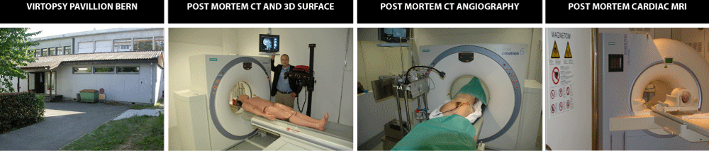
Our Forensic Imaging & Virtopsy® Courses provide theoretical and relevant practical knowledge of forensic imaging, notably post-mortem CT and MR, forensic anthropology, post-mortem angiography, surface scanning and 3D-photogrammetry, robot-guided biopsy, and clinical forensic imaging. For that, the course offers the participants computer access to actual cases that are then available for course/workshop training.
The courses are offered as a CAS (Certificate of Advanced Studies) in Forensic Imaging & Virtopsy. The certificate is issued by the University of Zurich and can be obtained after completing all assessments and course modules.
The modules can be taken separately, but not in any order.
- The Virtopsy® Basic course (module I) consists of two days with a focus on post mortem CT (PMCT). Lectures are followed by workshop sessions where participants will learn how to solve case related problems in context of the preceding lecture on a CT workstation. So a lecture about drowning will be followed by a hands-on session with cases where drowning was an issue.
- The Virtopsy® Advanced course (module II) takes three days, and it will make the imaging (PMCT, post mortem MRI) of a concise case that is followed by a full forensic autopsy available to course participants. There will also be a similar case demonstrated in full examination for PMCTA (post mortem CT angiography). Also, the Advanced course expands the subjects of the Basic course with more detail.
- The Virtopsy® Practical course (module III) then starts the day after module III finished, that is, it starts on a Saturday and finishes the following Wednesday (compulsory parts) or Thursday (for those that wish to stay to work on their case books). It teaches how to read PMCT more systematically, how to provide convincing written and image documentation for forensic use, it teaches practically how to perform PMCTA and also, how to perform 3D photogrammetry for surface documentation.
There is now one more course part that is offered, that is not required for the certificate, which focuses on how to use software for medical radiology image reading. That part is optional, any participant that aims for the certificate does not need to sit this. And to keep our naming of this part in line with the strictly regulated terminology and University of Zurich requirements for Certificates of Advanced Studies, it is not termed “module”.
Target audience
The CAS Forensic Imaging and Virtopsy® is aimed at all persons who are professionally interested or actively involved in forensic radiology and imaging, including forensic pathologists/medical examiners, radiologists, coroners, radiographers/radiology technicians, surveying specialists, and forensic anthropologists.
Accreditation
The course director and coordinator as well as many of the course presenters are members of the International Society of Forensic Radiology and Imaging (ISFRI). The ISFRI also works towards coordination across best practices, education, research, quality standards, certification as well as providing a journal for the members of the society, the Journal of Forensic Radiology and Imaging.
The course is submitted for accreditation to the EACCME every year. It also has been accredited by the ISFRI.
- The course modules I, II and III are compulsory parts for the certificate of the University of Zurich; these are awarded ECTS points by the University of Zurich that count towards the CAS Forensic Imaging and Virtopsy.
- We regularly apply for EACCME accreditation. Depending on the specific time schedule of the course, each course part (pre-course,1,2,3) is awarded a number of CME points. In 2019, the pre-course was awarded 6 CME points, module I was granted 13 CME credits, module II was granted 19 CME credits and for module III, 28 CME points were granted.
- We also applied for ISFRI accreditation which was approved in 2019.
Remarks about the course subject choices that we made
- The course also introduces new technologies such as 3D-printing, 3D-visualization, and Deep Learning. We have research in these areas and feel is relevant to show how these link into post-mortem CT-imaging.
- The 3D surface documentation session is an addition that is regarded as particularly relevant because of a recent rise in surface documentation also in forensic science where crime scenes and scenes of traffic accidents are increasingly documented in full 3D. An introductory lecture is provided in module I, and a full day of practical instruction is performed as part of module 3.
- Post mortem CT angiography is now taught and practically expanded on the Virtopsy® Practical course. Participants will also receive a full PMCTA kit (pump, power supply, materials, tubings, etc.) complimentary with the practical course.
- How to use the software will be a clear non-issue for radiologists but it can be relevant for forensic pathologists to learn this aspect in more depth. Please sign up separately for the Virtopsy® PRE-Course if you are interested.
Technical / skill requirements
Like the modules I and II of the CAS Forensic Imaging & Virtopsy®, fluent use of computer (such as Windows system) and English language are required.
Also, a medical background is clearly assumed as laid out in the statutes of the University regarding this course. Exceptionally, participants with other background may be admitted or sit in, by agreement, but this very clearly is a medical course aimed at people that practice or plan to practice forensic post mortem CT imaging.
Computer and language skills required:
- Proficient use of a computer (mouse, keyboard; Word, Powerpoint) as using software is a predominant substrate in imaging generally and in post mortem imaging specifically. If this is an issue please take a local software use course beforehand.
- English language skills that are sufficient to both understand others and express oneself both orally and in writing in an international setting.
Content of course parts
There is a Basic Course (2 days) and, immediately following, the Advanced Course (3 days) and a Practical Course (3,5 days, optionally up to 5 days). A 1-day Visualization Pre-Course has been defined to teach 3D viewing software to people that request this.
Course room setup and practical aspects of all course modules
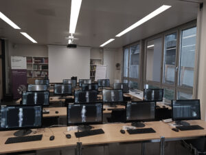
Many lectures of the course modules have actual cases, where participants examine the post mortem CT data themselves on the computer that is set up for every participant in the course room.
The practical aspect of investigating a post mortem CT are deemed relevant from our view and every year, we set up the course room accordingly. Course fees also cover the extensive server/hardware/sofwtare setup for this course.
Visualization Pre-Course (DICOM image manipulation and reading)
This facultative part is offered preceding the regular CAS Forensic Imaging Course modules.
It teaches the practical use of CT (or MRI) medical radiology image reading software including syngo.via (which is the system we have here for routine work and research, and that we also use for all three regular CAS Forensic Imaging and Virtopsy® Course modules) and also gives a short introduction for non-commercial DICOM reading softwares (3D slicer, Mevislab).
This is a full 1-day introduction into the technical aspects on how to use DICOM data manipulation and reading software for the benefit of applied forensic imaging.
As that, it provides relevant practical teaching on how to use axial image viewing, windowing, MPR (multiplanar reconstruction), and VRT (3D volume rendering technique).
It is not a pre-requisite for the regular course modules but it can make life a bit easier for participants that are new to forensic CT image reading.
It is available for registered participants of module 1 (and those that take modules 1 and 2, or 1-3) for a reduced price.
This is mainly geared towards the needs of forensic pathologists and other people participating in the modules I, and maybe II and III, that wish to learn basics of software use for CT data visualization.
While there are short software use introductions at the start of module I and also on the first day of module III, many participants over the years voiced their interest in learning software use in a greater depth. So we show how to use syngo.via (Siemens), and there is a relatively short introduction for 3D slicer and for Mevislab. This 1-day course takes place before the start of Virtopsy® Basic Course (module I).
Basic Course (module I)
This course covers general aspects of post mortem CT scanning. We introduce basic concepts of forensic imaging for pathologists and basic concepts of forensic medicine for radiologists. Reporting is a relevant topic for this course part where basic concepts are presented and discussed. A focus is on natural death, sharp force injury, blunt force injury, drowning, thermal injuries, and CT scanning protocols. We also explain basics for using post mortem CT for the confirmation of a presumed identity. The subject of how to set up one’s own CT scanning service from viewpoint of a forensic medicine facility is explained and discussed. Last but not the least, the use of forensic imaging for the benefit of clinical forensic cases is explained.
There is a special focus on Forensic Anthropology, as this field also is concerned with forensic post mortem imaging with issues such as commingled remains, mummy research, and mass disaster events. Particularly for forensic radiologists, this established area of examination and research is very relevant from our medicolegal viewpoint.
We may also include other new or relevant subject matter to present to the course participants as technology and research are evolving.
The course attendants will receive a Certificate of Attendance once they provided full attendance of the lectures of this course. A written assessment has to be passed. One ECTS point is awarded. We regularly apply for CME credits as well.
Advanced Course (module II)
This module contains a 1/2 day where – if possible – an autopsy case is followed, first by examining the post mortem CT by all participants and then seeing the autopsy findings demonstrated to the participants.
It furthermore contains a 1/2 day where – if possible – a post mortem CT angiography is performed, and findings then are examined by the course participants and discussed in detail.
Further topics cover strangulation, imaging of abuse and neglect, vital reactions, foreign bodies, technical subjects such as lung ventilation and CT-artifacts, post mortem MR. More research oriented topics include 3D data structures, a forensic view on the use of finite element modelling, magnetic resonance spectroscopy (1H-MRS) and basics of 3D surface documentation by photogrammetry.
Module 1 is a requirement for module 2. The course attendants will receive a Certificate of Attendance once they show previous attendance of module 1, and once they provided full attendance of the lectures of this course. A written assessment has to be passed. One ECTS point is awarded. We regularly apply for CME credits as well.
Practical Course (module III)
This module is geared towards actual users of post-mortem CT scanning and teaches practical forensic CT reading and documentation, we also teach CT angiography using very affordable materials, and practical 3D surface photogrammetry using consumer cameras. The target audience for module 3 therefore will most likely be people that are responsible for actually reading cases and generating forensic imaging reports. So, module I and II are a requirement, as this module III is only for those that did the Virtopsy® Basic and Advanced Courses in the past. In addition to attendance of the course, assignments and tests need to be passed. The course attendants will receive a Certificate of Attendance once they provided full attendance of the lectures of this course and passed attendance and assessments; prior attendance of the previous modules is a must. Maximally allowed participants are 12. Four ECTS point is awarded. We will apply this course for CME credits as well. The course is on-site here at the Zurich Institute of Forensic Medicine.
Module content distribution across modules I, II and III
We offer training so that participants ultimately can achieve a Certificate of Advanced Studies in the field of Forensic Imaging. The title of “Certificate of Advanced Studies in Forensic Imaging and Virtopsy®” is issued by the University of Zurich.
While the pre-course only covers software use skills, modules 1 and 2 are thought as general introduction and method demonstration, and to give participants workshop-like hands on experiences about various aspects. The module III however gets participants deep into practical use aspects, where own cases are examined in considerable numbers and participants, under guidance, build their own first visual documentations.
The module III of the CAS Forensic Imaging course is the main practical part, so we also call it “Virtopsy® Practical Course”. For anyone facing own application of the post mortem CT methods, this may be the most important part. The focus of module III is clearly on practical skills. Practical skills are taught in post mortem CT reading and report preparation, post mortem CT angiography and 3D surface documentation, and many cases are actually viewed by every participant themselves, during the workshops.
Module III – target audience
Students of the CAS Virtopsy® are expected to have a Master’s degree in medicine from an accredited university and certification as a specialist in forensic medicine or radiology. In exceptional cases, persons with a Bachelor’s degree in a medical field from an accredited university and several years of work experience in radiology, or persons with an equivalent qualification may be admitted to the program.
Module III will be most useful to all those that start their own post mortem CT scanning service and want to practically learn how to make best use of the data.
Practical focus of module III
While PMCT and PMCTA are widely published examination and scanning methods, we are convinced that there is a particular approach to this to make it work for daily forensic casework.
To help course participants to get the necessary traction, we, therefore, identified thorough PMCT reading, effective comprehensive written and graphical reporting, practically performing a PMCTA (post mortem CT angiography), and being able to practically perform 3D photogrammetry as key content.
Participants will be guided and taught through a set of cases and findings. They will learn and perform image reports both written and graphical. And there will be a practical PMCTA workshop with a newly developed very affordable PMCTA kit that contains all necessary materials for anyone to perform a PMCTA. Every participant will be equipped with such a kit.
Formal aspects
The CAS in Forensic Imaging of the University of Zürich, Switzerland, currently is the only public University degree in Forensic Imaging that is offered for non-profit. Details are outlined in Regulations Governing the Continuing Education Program CAS in Forensic Imaging and Virtopsy® at the Faculty of Medicine, University of Zurich, Switzerland [pdf]. Most notably, this University certificate can only be issued if certain requirements are met. Graduates of the full CAS Forensic Imaging and Virtopsy® will be awarded a total of 10 ECTS points. Requirements to be met are attendance of the course modules I and II and passing of the assessments there (both modules) (2 ECTS), the attendance of module III that contains the provision of practical project work (3 ECTS), and providing documentation for one real case in our institute under our supervision (1 ECTS). Last but not the least, a case book has to be submitted documenting 50 post mortem cases (4 ECTS). Time-wise, the total days of actual presence required at our institutes is 5 days (for modules I and II formerly called Virtopsy® Course, which is an integral part of the CAS, or a requirement) and another 3,5 (compulsory) but up to 5 days (last 1 1/2 days are facultative) for further course work of module III. The case book that the participants will prepare may be done off-site (for example, at their own workplace) and it has to be submitted to the CAS course coordinator. It is suggested to provide and contain 50 (fifty) single case studies from the own caseload of participants. The deadlines to submit the casebooks are given below.
Module III – program
The schedule for module III has been established as follows:

Module III – materials for PMCTA / set replacement parts
Module III also introduces participants to post mortem CT angiography. Every participant is issued with their own suitcase / kit with materials to perform a post mortem CT angiography; the kit contains a pump, power supply, tubes, and materials to cannulate the femoral vessels.
Module III – how to make a CAS casebook (50 cases)
Each participant has to prepare and submit a book containing 50 case descriptions. These then are assessed by the Virtopsy® team. The casebook is usually the last step in completing the studies necessary to obtain a CAS in Forensic Imaging & Virtopsy®. This does not have to be completed on-site – usually, participants make their casebooks from where they usually work.
The CAS for Forensic Imaging and Virtopsy® requires you to prepare a casebook containing 50 case studies. Based on both our own case-based experience and scientific studies into the effect of written and depicted reporting, we are convinced that PMCT evidence needs to be presented carefully to the attorneys or authorities that authorize you to perform a forensic PMCT on a body. The suggestions we make for your casebook base on our experience and insights. Preparing the CAS Forensic Imaging and Virtopsy® casebook may allow you to practice the preparation of PMCT evidence in a way that also authorities and courts can follow your descriptions.
- Make it a somewhat wide choice of forms of violence or trauma, and focus on positive findings
- 1 page per case is sufficient, >5 pages per case probably a bit much
- Images if necessary with support for orientation, i.e., small navigator overview or labels (i.e., left, right, ..)
- Labels/arrows to all findings that you provide written text/captions for, figure captions both descriptive and with meaningful interpretation or diagnosis
- Skeletal injury AND Vital reactions if applicable
- Conclusion / interpretation / what you think it is
Downloads:
Module III – reporting forensic PMCT cases
While we teach and mostly use straightforward tools (such as Microsoft Word and Powerpoint) to generate our written and visual reports, reports can have many forms.
Some reports that one may encounter seem quick and fast, where quickly generated visual output is made by simple procedures, some are exhausting to achieve and perfected in their appearance.
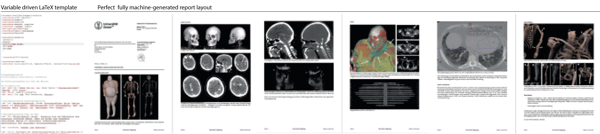
The manually optimized solution are commercial text- and image layout processing products; there, one stands on the shoulders of giants there: their optimization has been approximated through many, many software revisions (as examples, Microsoft Word has seen revision changes since 1983, that is, for around 35 years; Microsoft Powerpoint has been repeatedly revised since 1987, that is over 30 years, so in terms of pure manipulation efficiency, these are a hard act to follow). However, parameter-driven report generation may be interesting as an alternative, where one may work towards fully scripted, variable and ultimately database-driven report designs, where text, images, and captions obtain a perfected print layout without manual interference with the actual nudging, placement or alignment steps that often are so time-consuming. When using LaTeX possibly as middleware, the quality of the typesetting and postscript layout can be maximized in such a way – also because LaTeX has been repeatedly revised and improved since 1985, that is, 33 years.
Whatever the concise solution one chooses, the principles of forensic PMCT reporting remain the same – the client will require all relevant information, in an understandable and concise but not too abbreviated fashion.
Overview over a LaTeX template-based report with automated layout:
Links to some online-form check-list type assistance and an article to help to understand the issues:
- Short Form (English)
- Long Form (English)
- Article about a check-list approach to structured reporting
Visual case documentation
The visual (and not only written) case documentation is a focus subject for this Forensic Imaging course that is pursued particularly in module 3, so that all participants can actively learn the relevant details. At the latest then, putting together a good case book of 50 cases for the last assessment (see elsewhere on this page) should not pose technical obstacles any longer.
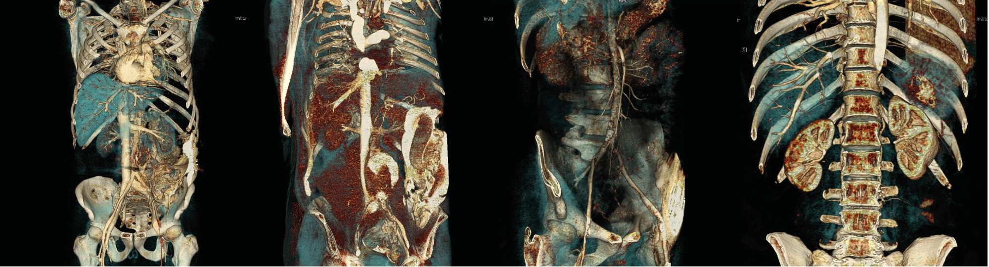
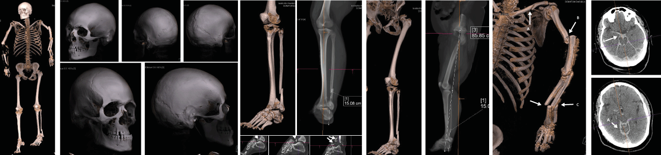
ex.: trauma case
A trauma case may be documented both in relation to skeletal injuries as well as organ or soft tissue trauma. The image (below) gives an overview of such a visual documentation.
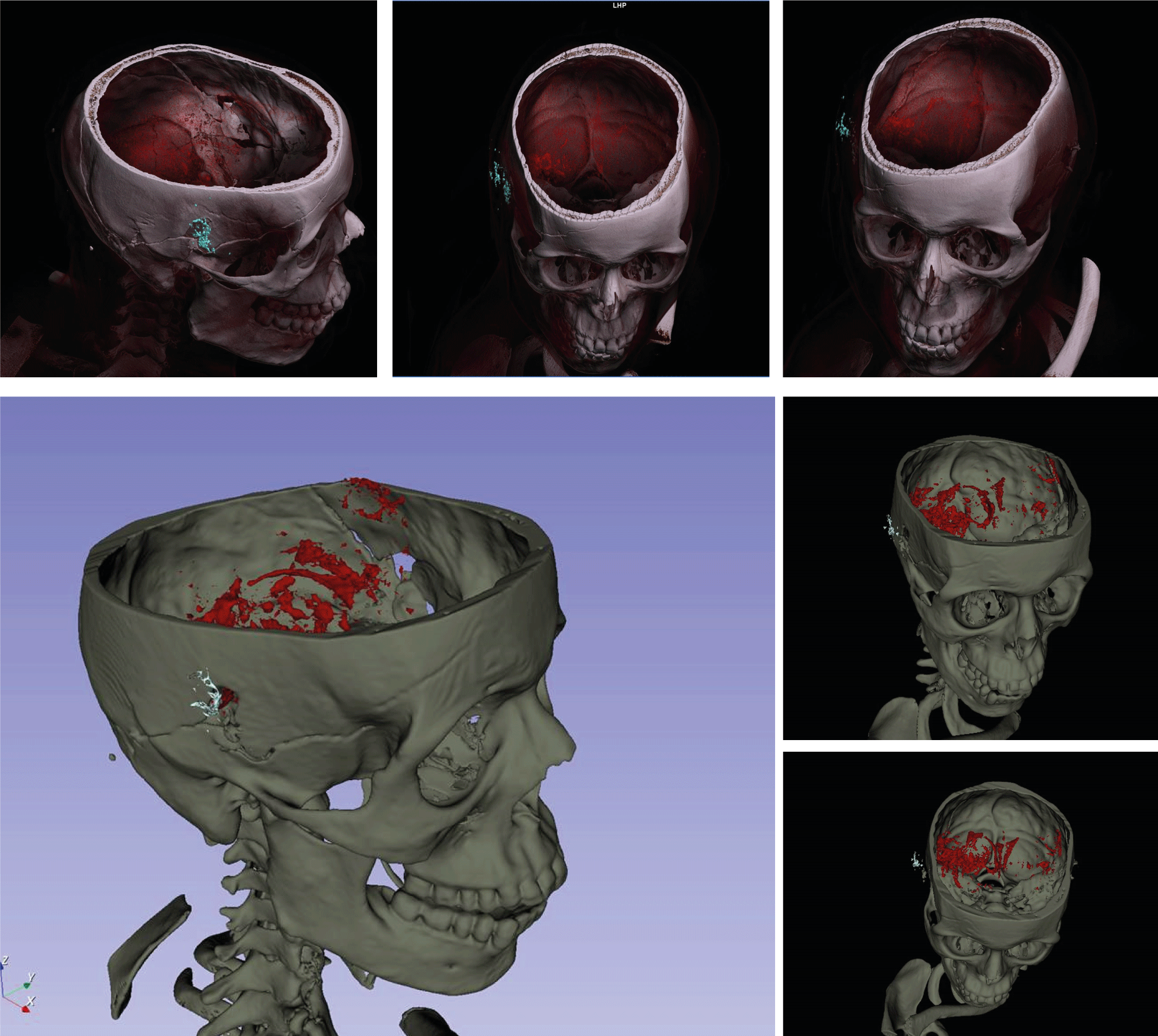
ex.: firearm injury to the head
In a case of close-range / contact head shot, subcutaneous findings at the entrance wound (cyan: ring-shaped CT-findings in subcutaneous tissue that are likely to be caused by gunshot residue), fracture pattern and intracranial findings (red: blood density range) can be examined for shape, extent, and distribution.
The following image shows Slicer 3D (below images: polygon segmentation, surface ready for STL-export for further applications such as rendering or 3D-printing) and syngo.via (Siemens) imagery (top three figures: VRT reconstructions after segmentation).
The single steps involved in axial, MPR and also VRT viewing using syngo.via-Siemens software as well as steps in segmenting in Slicer 3D are taught in the facultative Virtopsy® Pre-Course.
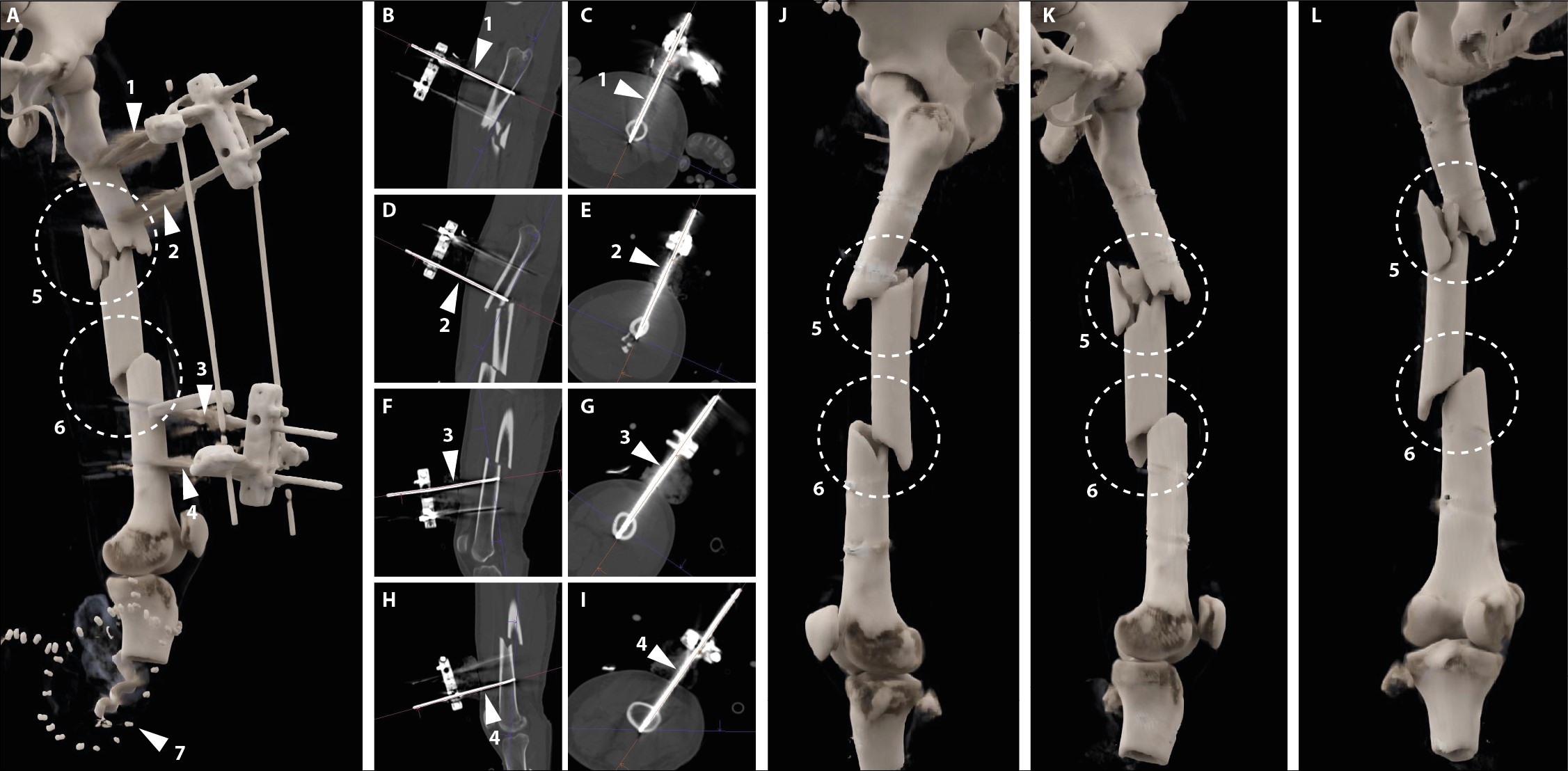
ex.: metal implants
Metal implants – such as a fixateur externe – may be visualised in detail as shown in the following image. The rods and details of the fracture fragments are demonstrated.
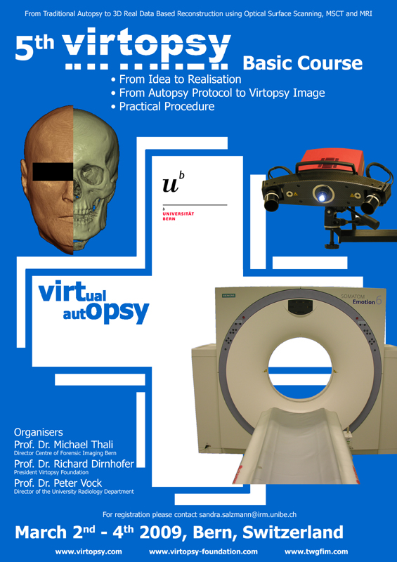
Past courses
Certificate for Advanced Studies Courses
Course 2017
- March 20th-30th (Modules I-III)
- Deadlines for casebook submission I: 30th June 2017, II: 31st December 2017; III: 30th March 2018, IV: 30th June 2019. Last: five years after, 2022.
Course 2018
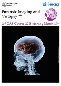
- March 19th-29th (Modules I-III)
- Deadlines for casebook submission I: 30th June 2018, II: 31st December 2018; III: 30th March 2019 (1 year after course end), IV: June 30th 2019. Last: 2023, five years after.


Course 2019
- March 15th (Pre-Course) / March 18th-28th (Modules I-III)
- Deadlines for casebook submission I: 30th June 2019, II: 31st December 2019; III: 28th March 2020 (1 year after the course end), IV: June 30th 2020. Last: 2024, five years after.
Course 2020
- Cancelled on first day of module I due to COVID-19 related lockdown.
Course 2021
- Not conducted.
Course 2022
- March 11 (Pre-Course) / March 14-24 (Modules I-III).
- Deadline for casebook submissions: every June 3oth and December 31st, until five years after the course which will be 2027.
Course 2023
- March 7 (Pre-Course) / March 1119 (Modules I-III).
- Deadline for casebook submissions: every June 3oth and December 31st, until five years after the course which will be 2028.
Course 2024
- March 3 (Pre-Course) / March 6-22(Modules I-III).
- Deadline for casebook submissions: every June 30th and December 31st, until five years after the course which will be 2029.
Course 2025
- March 7 (Pre-Course) / March 10-21(Modules I-III).
- Deadline for casebook submissions: every June 30th and December 31st, until five years after the course which will be 2030.
Original Virtopsy® Courses
2011 7 March 8 November
2012 9 March 2013 10 March1 2014 11 March2
2015 12 March 2016 13 March 2017 14 March CAS 2018 15 March CAS
2019 16 2020 2021 (travel restrictions, no courses) 2022 17 2023 18 2024 19 2025 20 2026 21