About Virtopsy
Learning Virtopsy
Forensic Imaging and Virtopsy can be learned by self practice, reading and participating in trainings and courses.
Our course information is available here.
Contact Virtopsy

You may contact the Virtopsy office through E-mail – virtopsy@irm.uzh.ch.
What is Virtopsy
Today, computed tomography as a main focus, as well as magnetic resonance imaging and spectroscopy, optical 3D surface scanning, and 3D photogrammetry may routinely be used to search for and document forensic evidence in a non-invasive or minimally-invasive and observer-independent manner in both the living and the deceased.

Virtopsy began at the turn of the millennium as a multi-disciplinary applied research project to implement imaging modalities from diagnostic radiology and surveying technology in the routine workflow of forensic medicine [link]. Since then, the Virtopsy approach has become an emerging if not standard procedure in forensic investigations worldwide. The term Virtopsy has been used in a variety of settings all over the world (check Pubmed).
The methods are undergoing further research, validation and development; see the following examples or check our publications [link].

Virtopsy workflow. From: Ebert, L. C., et al. “Forensic 3D surface documentation at the institute of forensic medicine in Zurich–workflow and communication pipeline.” Journal of Forensic Radiology and Imaging 5 (2016): 1-7. [link] |

3D reconstruction of femur fracture. From: Flach, Patricia M., et al. “Imaging in forensic radiology: an illustrated guide for postmortem computed tomography technique and protocols.” Forensic Science, Medicine, and Pathology 10.4 (2014): 583-606. [link] |

Skull plane definition. From: Ruder, Thomas D., et al. “Comparative radiologic identification with CT images of paranasal sinuses–Development of a standardized approach.” Journal of Forensic Radiology and Imaging 7 (2016): 1-9. [link] |

Aortoesophageal fistula in Post Mortem CT Angiography (PMCTA), both using VRT (left) and Cinematic Rendering (right). From: Ebert, Lars C., et al. “Forensic 3D visualization of CT data using cinematic volume rendering: a preliminary study.” American Journal of Roentgenology 208.2 (2017): 233-240. [link] |

Myocardial infarction in MRI. From: Crooijmans, Hendrikus JA, et al. “Feasibility of post mortem cardiac proton density-weighted fast field echo imaging in two cases of sudden death.” Legal Medicine 15.6 (2013): 310-314. [link] |

Bar-chart detailing post imaging levels of confidence in relation to post-imaging categories of causes of death. From: Ampanozi, Garyfalia, et al. “Accuracy of non-contrast PMCT for determining the cause of death.” Forensic Science, Medicine and Pathology (2017): 1-9. [link] |
Quality management: ISO 9001:2008 certification
 |
The Departement of Forensic Imaging and Medicine of the Institute of Forensic Medicine of the University of Zurich has obtained quality management certification to ISO 9001:2008 for all three domains clinical, post mortem and radiological examination, including reporting and written expert opinions. |
How can I learn how to use concepts related to Virtopsy?

Ultimately, Virtopsy aims at improving your case investigation, decision making, and documentation quality. This means that scanning techniques have to become integrated with your workflow in a supportive way.
- You could visit one of our Virtopsy courses (and if you are interested in extensive training, continue with the CAS course) where participants have the opportunity to learn state of the art theory and practice from us, here, where Virtopsy started.
- You could read our publications or watch informative movies about Virtopsy.
- You could join the ISFRI, attend annual conferences and get the society’s journal, the Journal of Forensic Radiology and Imaging (JOFRI): the International Society for Forensic Radiology and Imaging (ISFRI) was founded to promote international information exchange and communication within the field of forensic imaging.
Virtopsy in forensic death investigations
Virtopsy has the capacity to enhance traditional autopsy or even obviate it in selected cases. One of the main benefits of imaging lies in an observer-independent documentation of many forensically relevant findings. Digital imaging data can technically be stored permanently and maybe re-examined at any time if a second opinion is required.Limitations are method inherent, and on the result side they relate to the limited resolution both of contrast and detail, and of specific limitations in both specificity and sensitivity whereas on the effort side, limitations are usually time and cost.
Virtopsy in cases of living patients
In living patients, Virtopsy permits the documentation of injuries such as bite marks, bruises, lacerations, and abrasions. Documentation may be approached in three dimensions, true to scale, and enables comparison particularly of patterned injuries to potential injury-causing instruments.
Virtopsy in court
Virtopsy has the capacity to provide excellent tools for death investigation, crime and accident reconstruction, including 3D depictions of internal injuries, 3D true color representations of surface injuries and even 3D scaled models of entire crime scenes and events. The Virtopsy approach has the capacity to document and present critical forensic evidence in an objective and comprehensible fashion, also suitable for presentation as evidence to laypersons and legal professionals.
Equipment
Virtobot

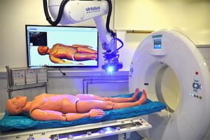
The Virtobot system is a robotic system that performs a variety of tasks in conjunction with the CT scanner which was developed by the virtopsy team in collaboration with the Austrian Center for Medical Innovation and Technology (www.acmit.at). It allows for automated, high-resolution 3D surface documentation as well as CT guided post-mortem tissue sampling. Its modular design makes it easy to extend the system by adding more functionality in the future.
CT (Computed Tomography)

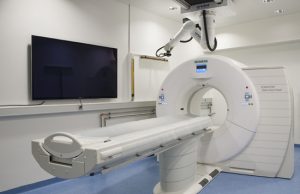
We are using a Siemens Dual Source SOMATOM® Definition Flash 2×128-slice scanner with a maximum scan speed of about 46 cm per second. Its Dual-Energy mode allows for a refined postmortem CT-Angiography (pmCTA), new post-processing techniques like artifact reduction and detection of material-specific differences. For visualization, a range of image reconstructions including MPR and 3D techniques are available on a variety of platforms.
MRI (Magnetic Resonance Imaging)

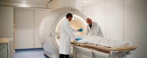
We use a 3 Tesla MRI scanner (Philips Achieva 3.0T). It covers both routine such as head or whole-body imaging and more sophisticated exams like MR-spectroscopy and diffusion tensor imaging (DTI).
Awards
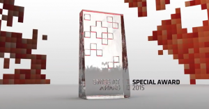
The Virtopsy Team has been awarded the Swiss ICT Special Award 2015.

